Orringer Lab

Research

The spatiality of glioma

We are studying early gliomagenesis to understand why gliomas form more often in some brain regions than others. When we mapped 204 IDH-mutant gliomas on MRI, we found 103 in the superior or middle frontal gyrus, about three times as many as expected from the volume of those regions.

Park et al., Neurosurgery
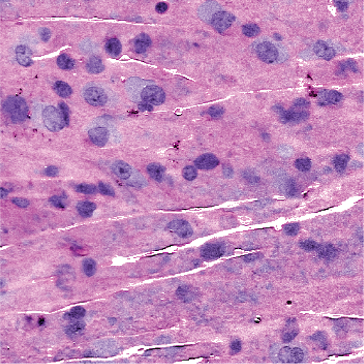
Imaging the tumor margin

Glioma cells can extend into tissue that looks normal to the eye. We use stimulated Raman histology to image fresh specimens without staining them, allowing for real-time intraoperative decision making based on data which was never before available.

Kondepudi et al., Nature
Low-grade glioma imaged with stimulated Raman histology
Low-grade glioma · Jiang et al., OpenSRH (2022)

Measuring tumor DNA during surgery

With the Evrony Lab, we developed a droplet digital PCR test that measures tumor mutations in surgical tissue in 15 minutes.

Murphy et al., Med